Many learners express a desire to deepen their understanding of anatomy and the body. You might recall passing your anatomy exam and thinking, “I don’t need to remember everything; I know how to teach a client to squat!” However, when unexpected challenges arise or clients move in ways that puzzle us, we often wish we could recall our anatomy lessons better.
As a kinesthetic learner and an educator for the past 16 years, I understand the difficulty of teaching complex subjects like anatomy. The key is to provide techniques that help learners anchor each body part to a memorable reference.
This blog series aims to offer movement practitioners a systematic approach to remembering anatomy, making it relevant and applicable in practice. I’ll share insights and techniques, including word association, history, and tactics, to make anatomy recall useful for training clients.
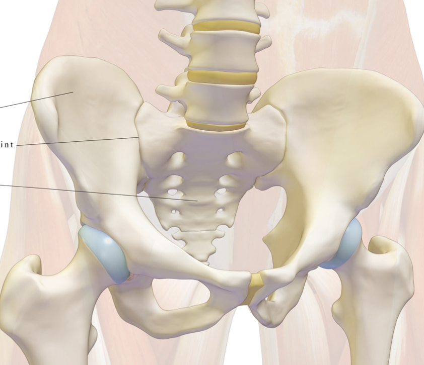
Anatomy books and skeleton models only provide a limited view of the pelvis. Observing real pelves reveals the structural diversity among individuals. Here’s a closer look at the key bones and their meanings:
 Key Bony Landmarks
 Understanding genetic differences in pelvic structure is crucial for tailoring exercise programmes:

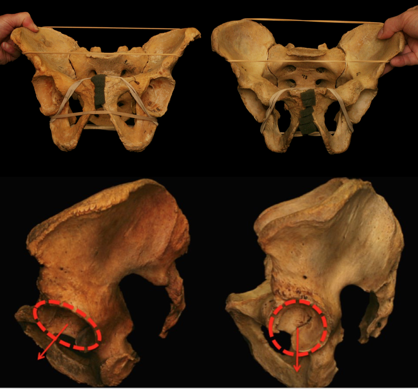
We know we genetically differ and as you can see here we can differ inside our bodies as much as we do on the outside but what does that mean to us as trainers?
Coxa Valgus or Vara & Femoral Version are good examples of terminologies for clients pelvis/hip geometry that differ in a way that may have clinical or movement relevance for their practitioner.
These can be identified using simple manual tests that we offer on our Running Biomechanics Workshops for example. They can change the way we prescribe exercises or what we expect of our clients when they move. Hopefully these descriptions help to appreciate this….
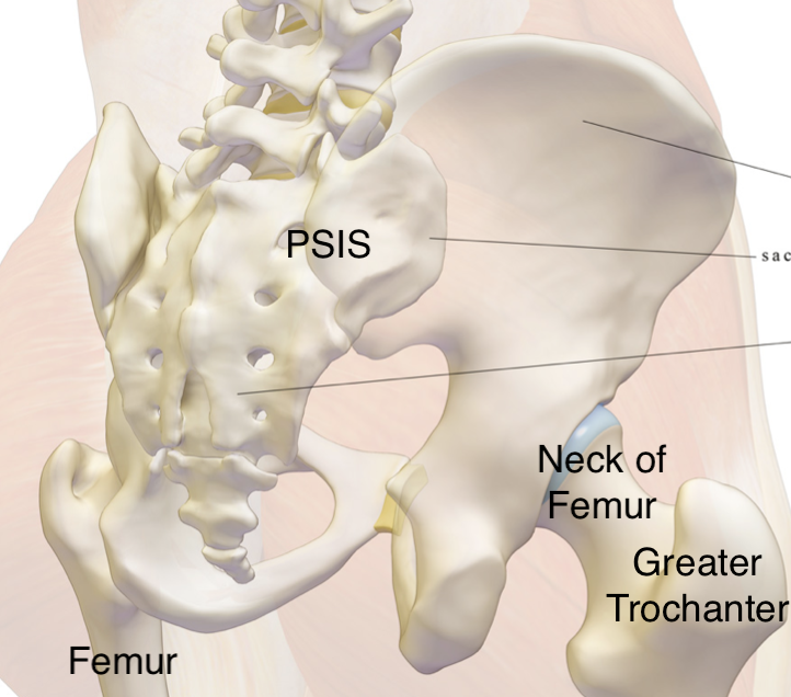
The Femur is the largest bone in the body & we will only discuss here the top (proximal) portion that relates to the pelvis. The other end (distal) will be discussed in the Knee Series.
Trochanter comes from the Greek word trokhos for wheel. It describes the large round feeling bony prominence at the side of the pelvis. It is a key landing site for the lateral glutes & piriformis – to be discussed next time.
Neck of femur is the part of the femur that looks like a neck sticking out from the medial side of the main femur thigh bone where the greater trochanter is lateral & then ends in the ‘ball’ of the hip’s ball & socket joint.
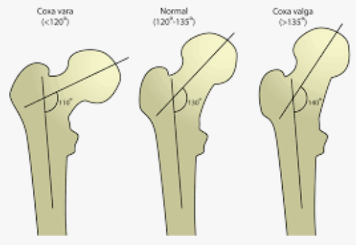
Coxa is a Latin word meaning hip or limb joint (koks-h)
Coxa Vara: A decreased angle at the hip, potentially limiting abduction.
Coxa Valga: An increased angle at the hip, potentially limiting adduction.
Imagine you have a client who has been diagnosed as Coxa Valga….how might this alter your expectations when you write them a programme or train them in your facility?
Femoral is related to the thigh or femur.
Version or Vers is Latin for turned or to turn.
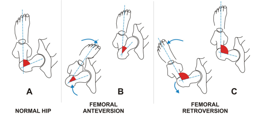
Femoral Version describes the rotation (of turn) of the femur in relation to the pelvis.
Femoral Anteversion: Medial rotation of the femur.
Femoral Retroversion: Lateral rotation of the femur.
Imagine you have a client who has been diagnosed as femoral anteversion….how might this alter your expectations when you write them a squat programme or train them in your facility?
Application in Training
Knowing these anatomical details helps explain why clients move differently. This knowledge is vital for designing effective training programs and assessing pelvic function.
Discover more with our Diploma in Biomechanics Coaching here