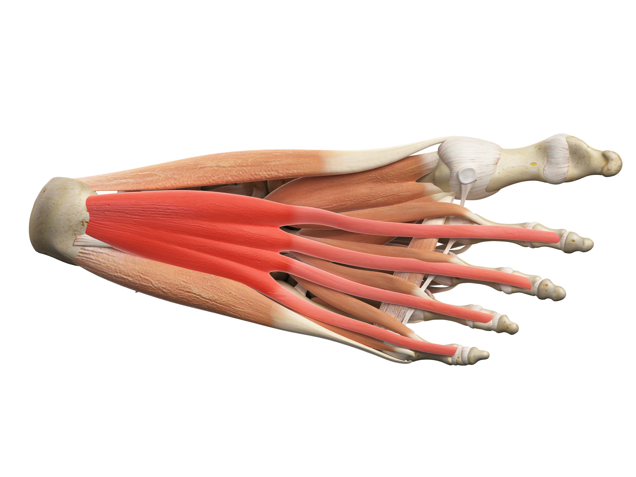


Shall we explore the final piece in this series to see if we can recall muscles in the foot and ankle to deepen our understanding of foot mechanics?
A fascinating finisher for you…
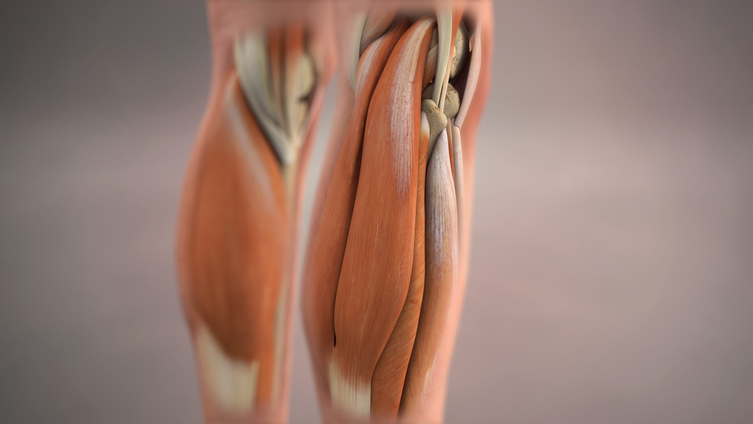
Gastrocnemius is also called a ‘calf’ muscle because it forms the large ‘belly’ of the leg & those Greeks thought it resembled a calf’s stomach/belly
Coming from the Greek words
gaster = belly
kneme = leg
It is the superficial of the 2 calf muscles with 2 heads that attach to the condyles (nobly bits) on the back of the femur. It also travels down to a tendon that becomes part of the Achilles (calcaneal) tendon at the back of the ankle/heel.
So for this reason, when it contracts or shortens it will plant the toes down into plantar flexion which is also what happens when you stand on tiptoe.
However if you wanted to lengthen it fully you would need to, not only dorsiflex the ankle (pull the toes upwards), but also extend your knee to take up the slack there too! Try this by taking one heel back behind you, with the toes pointing forwards, knee straight and heel down…you should feel a stretch in the back of your leg through the calf. In fact for many this could feel REEEEAAAAAAALLLLY tight!
Check out all the different ‘Gastrocy’ shapes out there next time you are around lots of bare legs or people in shorts or leggings!
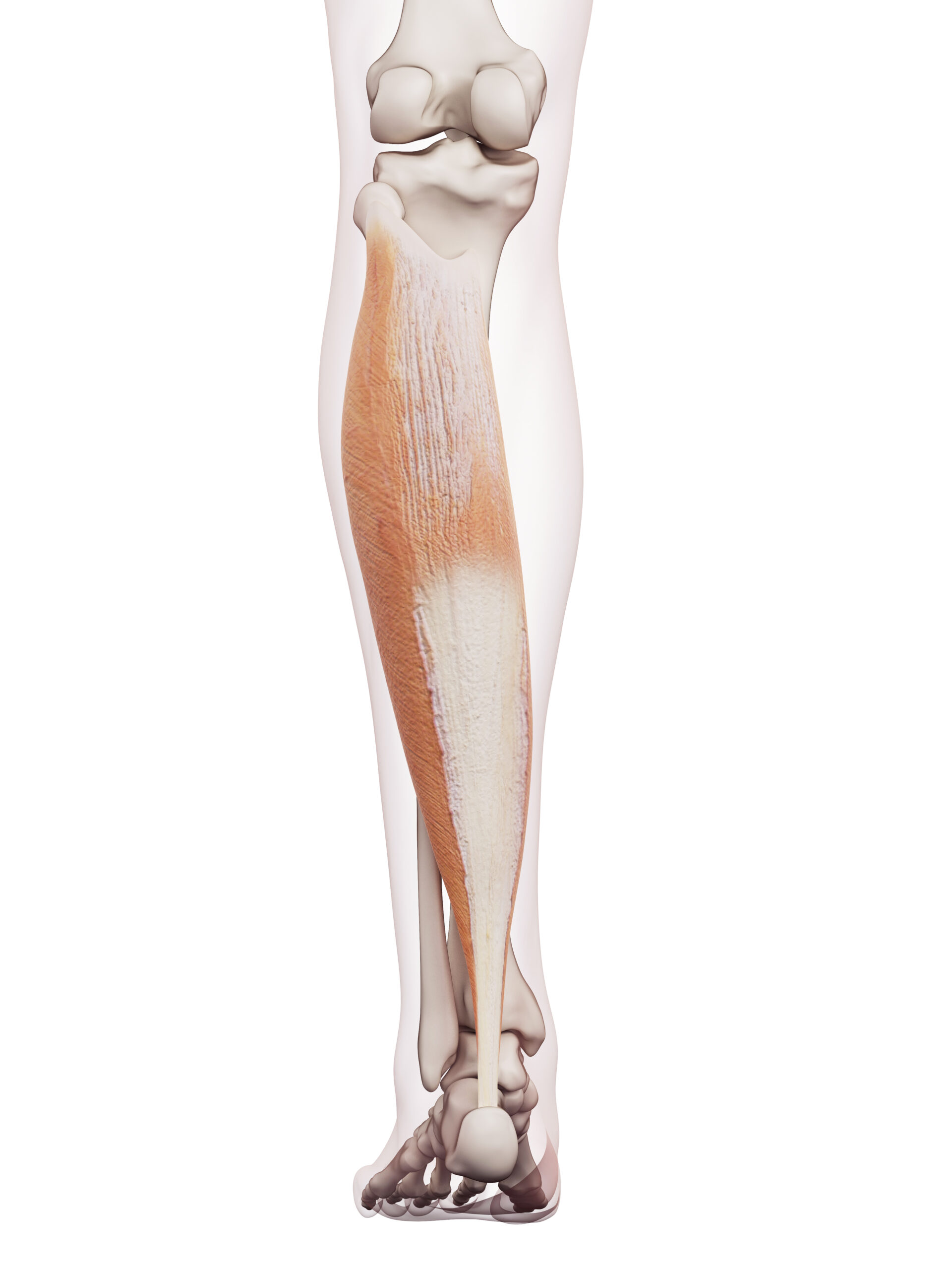
Soleus is the Latin word for a flat sort of sandal.
Deep to (underneath) gastrocnemius, this massive ‘pumper’ has an important job because it acts to pump blood back up the body, against gravity. Another great reason to suggest to our clients why walking at least 10k steps daily might be a good idea!
Soleus is also bigger than you think because much of its anatomy is hidden behind the gastrocnemius. It may well be flatter than gastroc but it’s muscle portion is certainly longer!
Starting on the back of the tibia & fibula it then travels down to the calcaneal (Achilles) tendon, onto the calcaneus (heel bone).
It is also responsible for pointing your toe like it’s partner, Gastrocnemius BUT Soleus can be lengthened or stretched even when the knee is flexed due to its anatomy NOT crossing the knee joint!
So if you wanted to see how it compares with the previous gastrocnemius stretch try doing what you did before…..place your heel down behind your midline, toe pointing forwards but this time, bend the knee and lower the body, keeping the heel on the floor. You should feel the Soleus stretch a little lower down.
You could encourage a client to feel both variations too!
Gastrocnemius & Soleus are also known as ‘triceps surae’ because it means 3 headed muscles.
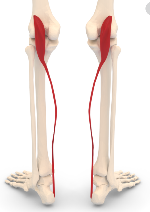
This slender muscle is considered unimportant or an ‘accessory’ muscle…
What is an accessory muscle?
An accessory muscle provides assistance in movement but isn’t classed as primarily responsible for it.
When it comes to the Plantaris muscle 8-12% of us don’t have one! (or 7-20% depending which article you read)
So by that calculation 88-92% of us DO have one which in MY book makes it a valid muscle
What do you think?
Sweeping from the outside of the lateral femur, across the back of the knee & down to the Achilles’ tendon, (also known as tendo calcaneus) it has the longest tendon in the body!
Whatever your opinion about Plantaris it flexes the knee & plants the foot down (plantar flexion)
A super short muscle belly that looks like a little rope, it also assists in allowing you to stand on tiptoes to reach up high into a cupboard
Go Plantaris!
Also known as the Peroneals OR Peroneus Longus & Brevis even!
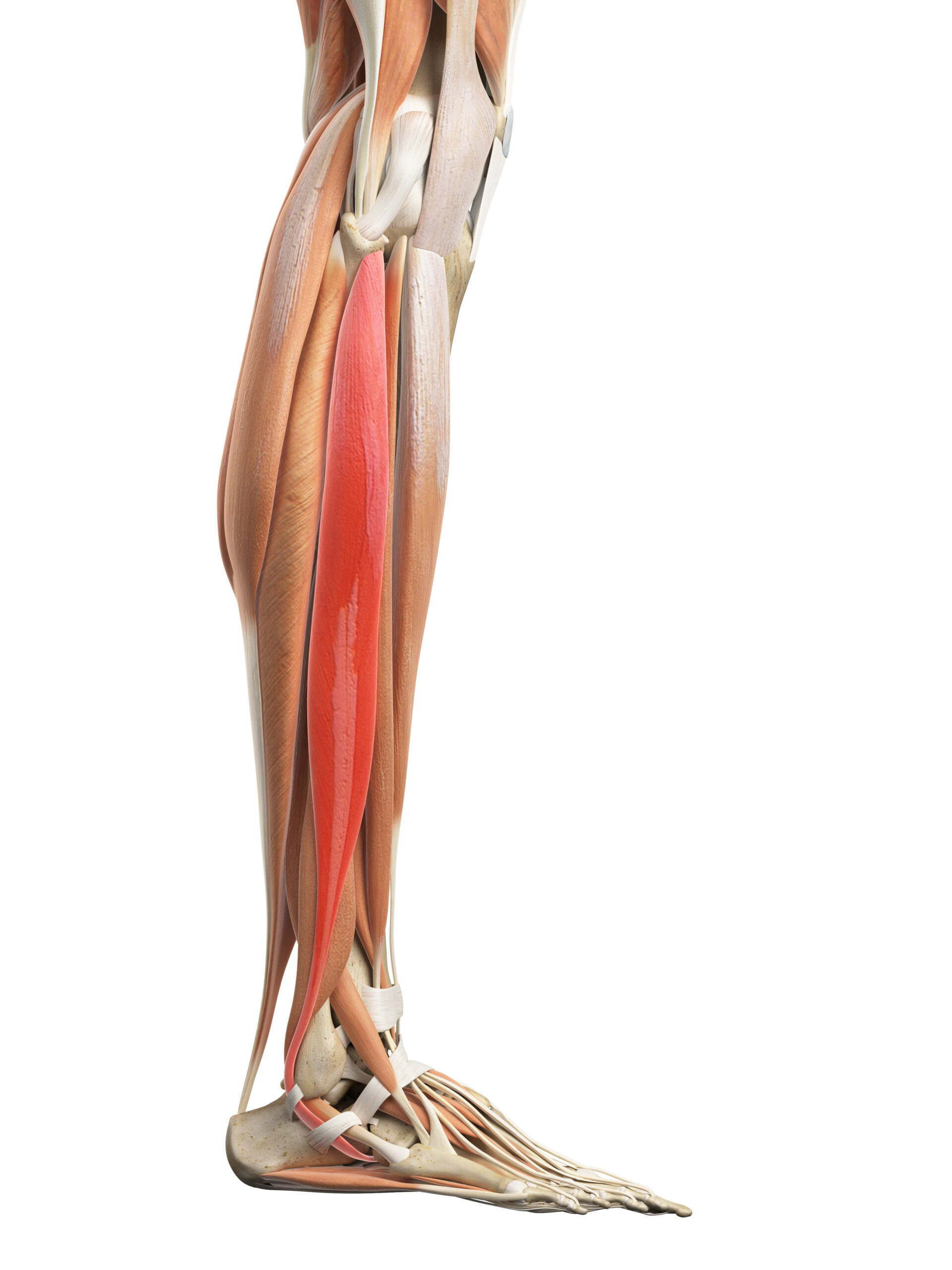
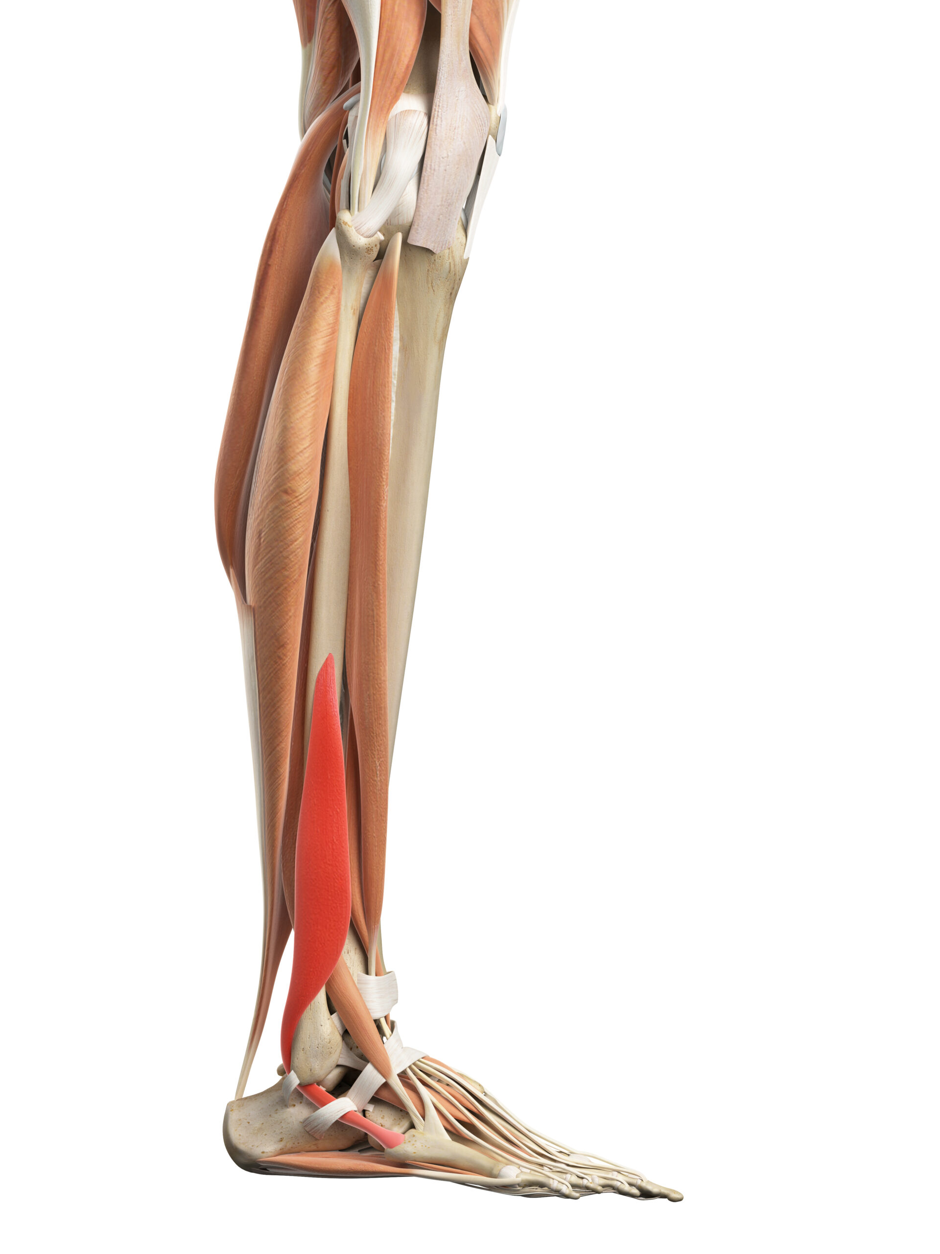
Peroneals comes from the Greek word Perone (pronounced Pair-uh-knee) which was their word for the pin of a brooch or a buckle.
Why?
Because the Greeks felt that the fibula looked like a pin at the back of the ‘tibia’ broach!
How to remember? Place your hands on the side of your lower legs, slide them upwards (over your perennials) towards your ‘pair uh knees’!
However these days they are being called the Fibularis muscles which should be even easier to remember since thats where your fibula is! It depends what resonates in your memory I guess!
Fibularis Longus is the long one, Fibularis Brevis the shorter one.
Fibularis Longus attaches from the proximal (upper) portion of the fibular & Fibularis Brevis joins in from the distal (lower) portion to travel down to the ‘plantar’ surfaces of certain tarsal & metatarsal bones, meaning they play a role in the movement of the ankle.
Fibularis Longus is a plantar flexor & everts the foot – meaning it turns the sole outwards. Through its tendon it also supports the arch of the foot…..so really multi tasking.
Fibularis Brevis helps his brother out through everting the foot too
Interesting fact: Fibularis Longus contributes to BOTH pronation & supination!
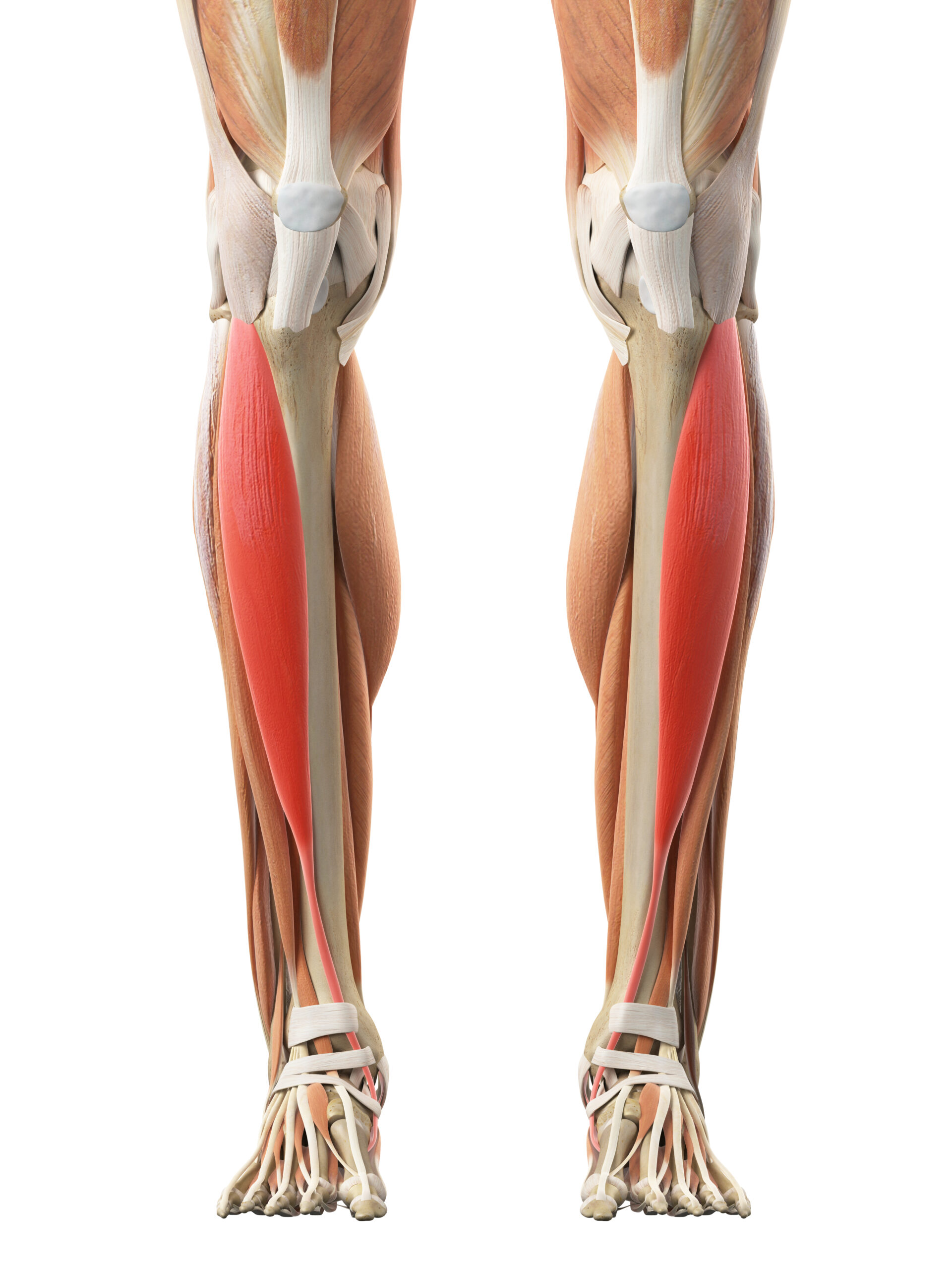
This bruiser sits on the front (anterior) of your tibia, hence tibialis anterior!
From the top lateral nobly bit to medial (inside) & underneath the first cuneiform, a bone that makes up part of the arch of the foot…..
This means it is able to lift, or dorsiflex, the forefoot. It also inverts (turns in the underside of the foot) & adducts the forefoot towards the midline.
So it should allow you to sustain an arch in your foot. The arch is important for supination, or ‘toe off’ when walking as it gives rigidity by ‘locking’ the midtarsal joints
Supination is the propulsion phase when walking
Tibialis Anterior also eccentrically decelerates your foot as you heel strike while pronated. It is often inhibited & an antagonist to those ‘tight’ calves. Or maybe it’s the Tibialis Anterior that inhibits those calves? Extends, dorsiflexes & everts by attaching to the lateral tibia to medial cuneiform & base of 5th metatarsal
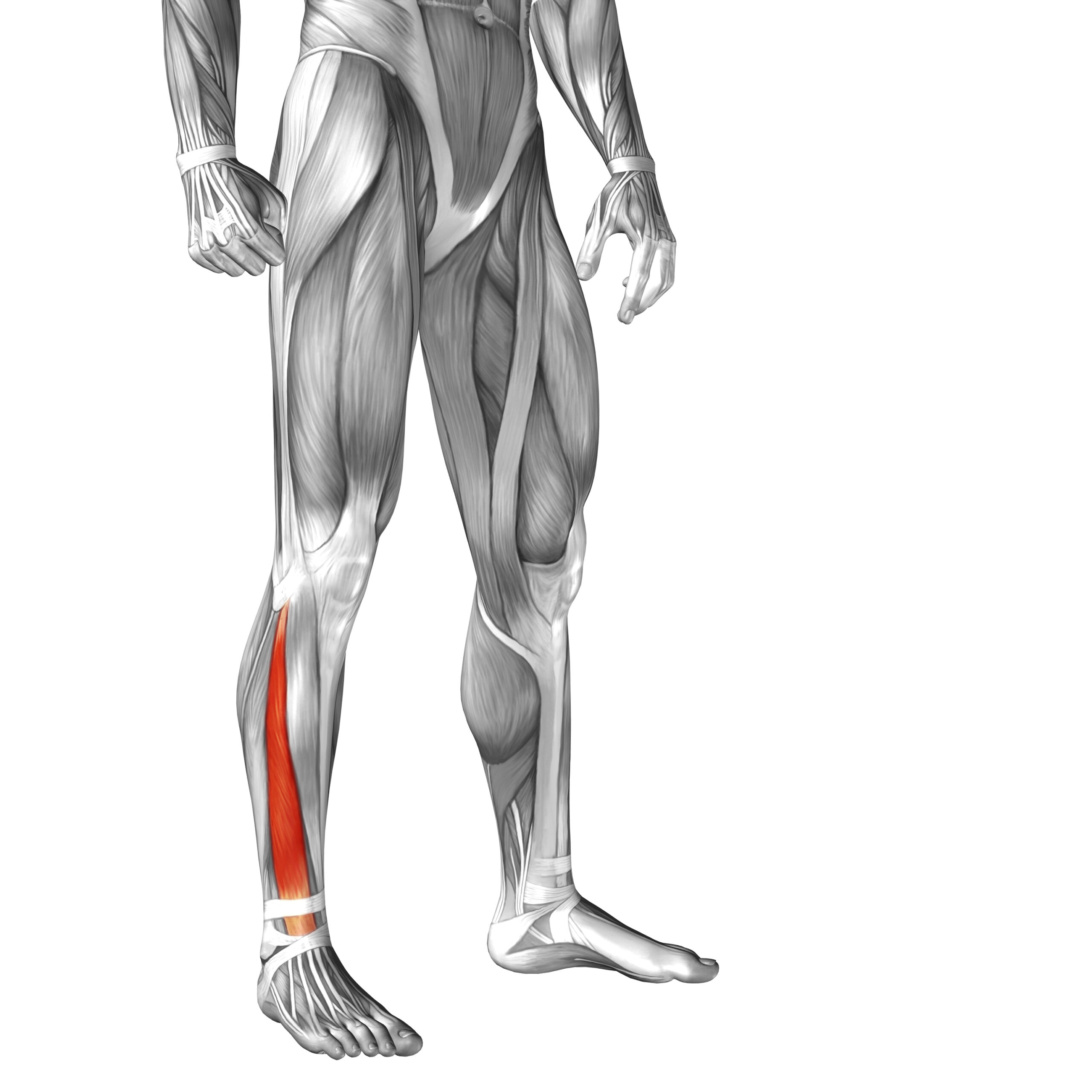
It extends the digits & is long…
In fact it extends 2-5th toes, dorsiflexes ankle & everts foot by attaching from the medial anterior fibula to phalanx of the first toe
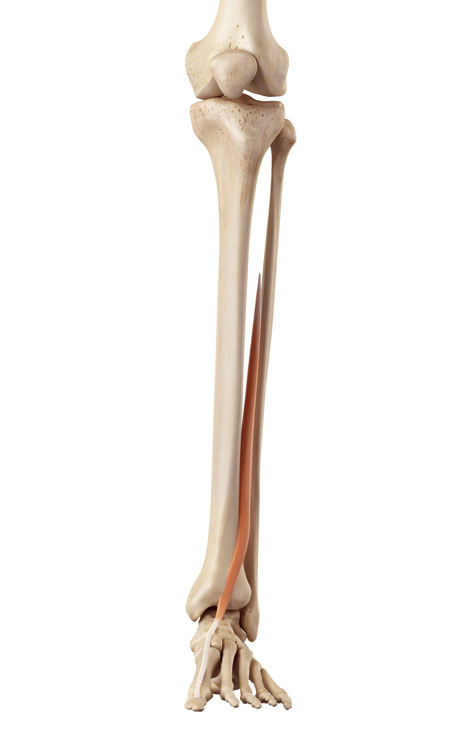
It extends the hallux & is long….
So it extends first toe, dorsiflexes ankle & inverts foot by attaching from the lateral condyle of the tibia to middle phalanges of 2-5th toe
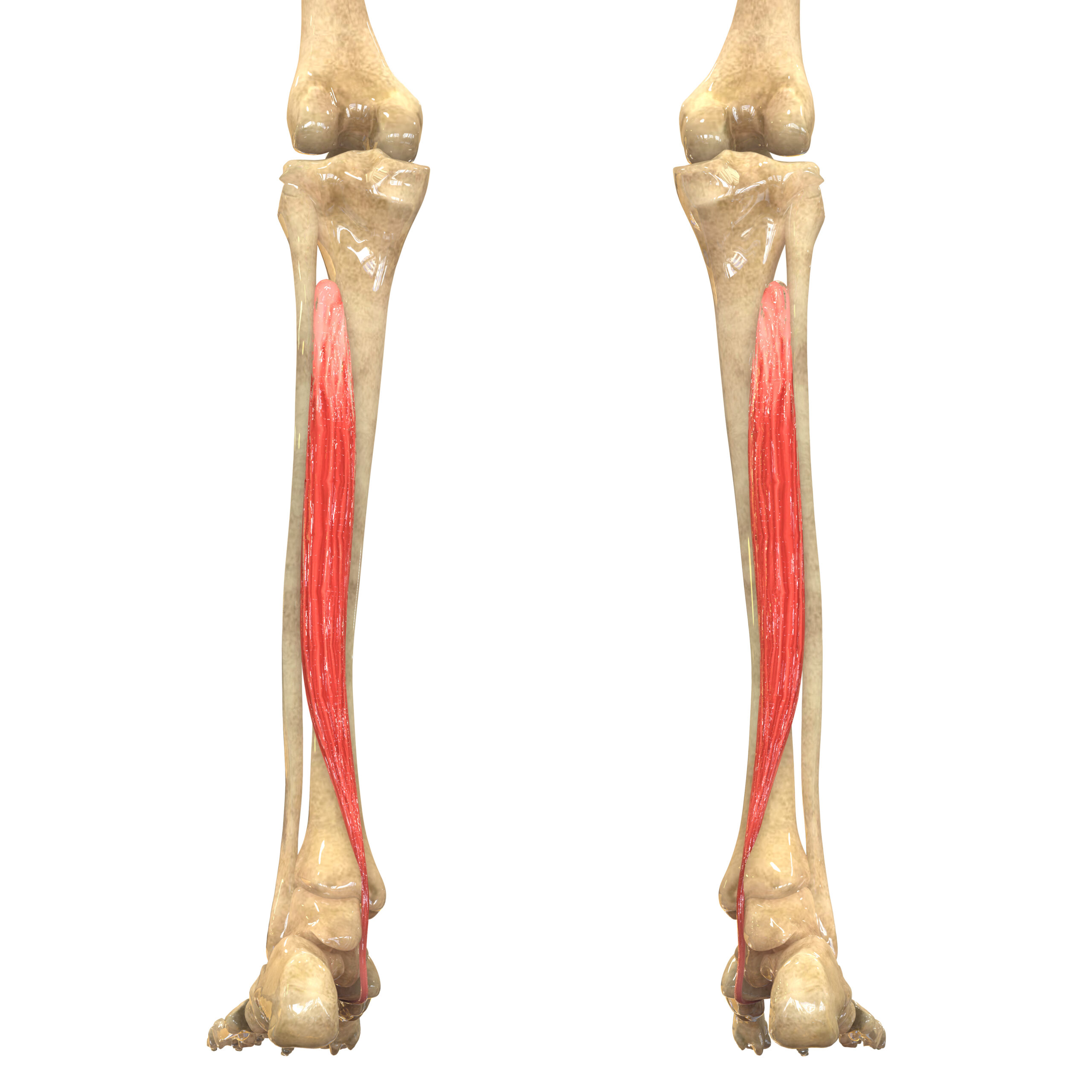
So this dude is in the BACK of the lower leg…yup you guessed it…posterior to (behind) the tibia.
A stabilising muscle that supports the arch of the foot, it can do this because it attaches from the upper lateral part of the tibia & upper medial part of the fibula….
Then inserts down onto the navicular (meaning ‘little ship’ as this bone is boat shaped) & medial cuneiform (Latin for ‘wedge-shaped’)
Takes care of plantar (planting down to the ground) flexion, along with its calf buddies, & inversion of the sole….
Therefore also a potential contributor to adults presenting with flat feet when dysfunctional or inhibited….
… by what?
Think 🤔 antagonistic… maybe!?!
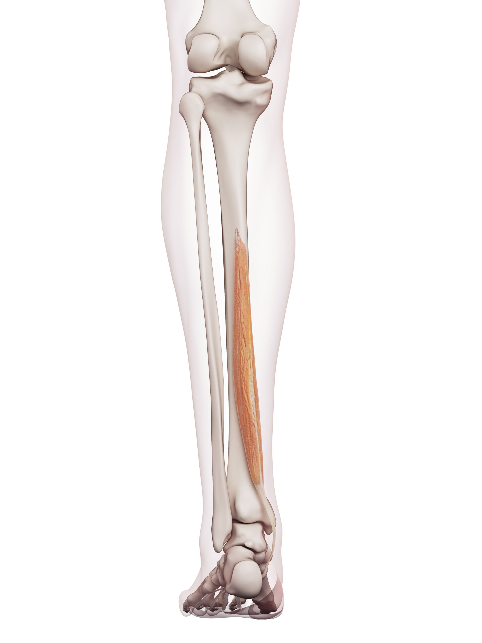
Flexes the 2nd-5th toes & is a weak plantar flexor of the ankle while inverting the sole of the foot by attaching from middle posterior tibia to phalanges of 2-5th toes
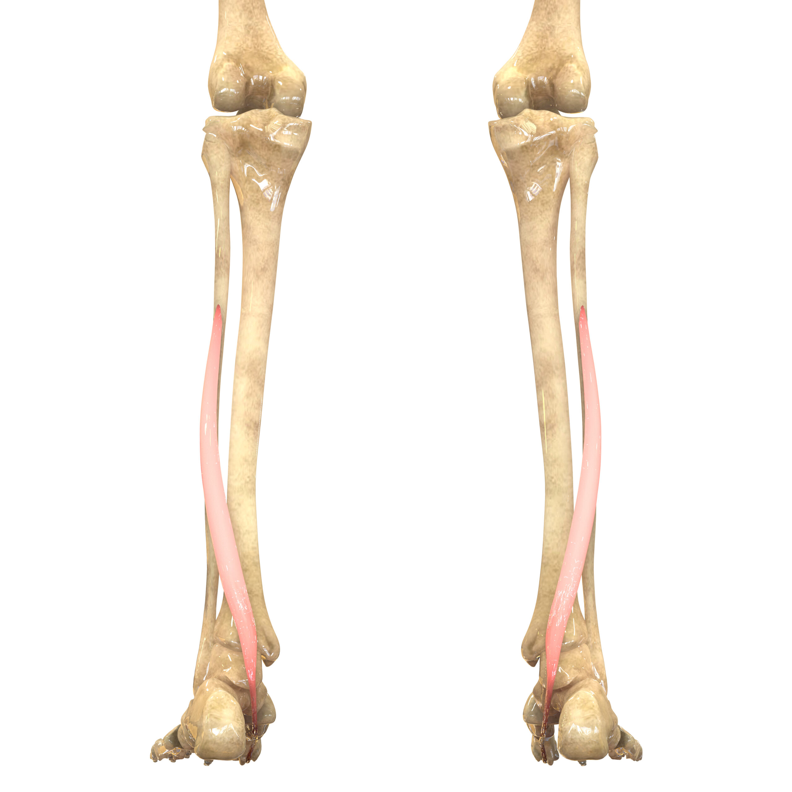
Flexes the Hallux or first toe & another weak plantar of ankle while inverting the foot by attaching to the middle posterior fibula to phalanx of first toe
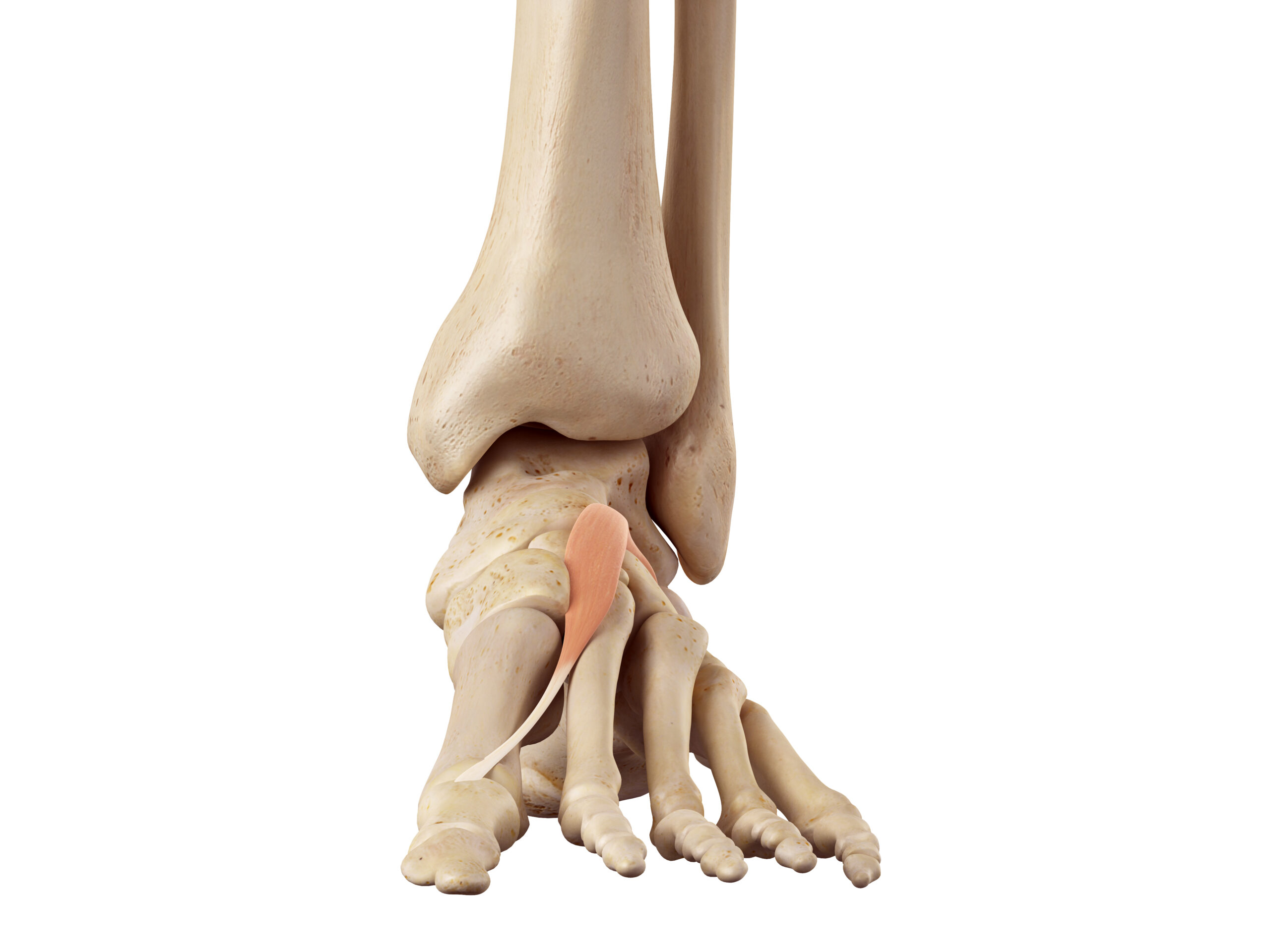
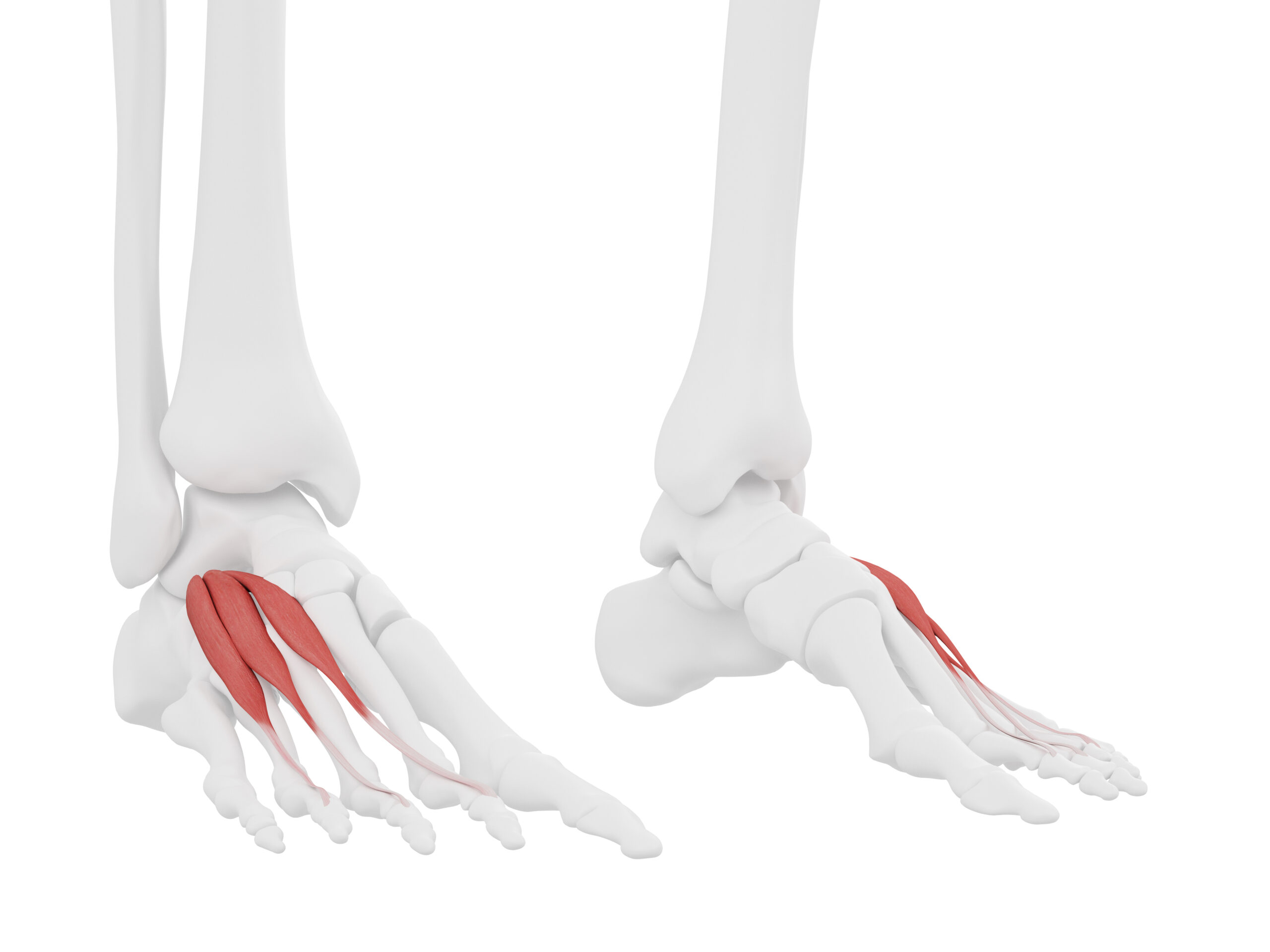
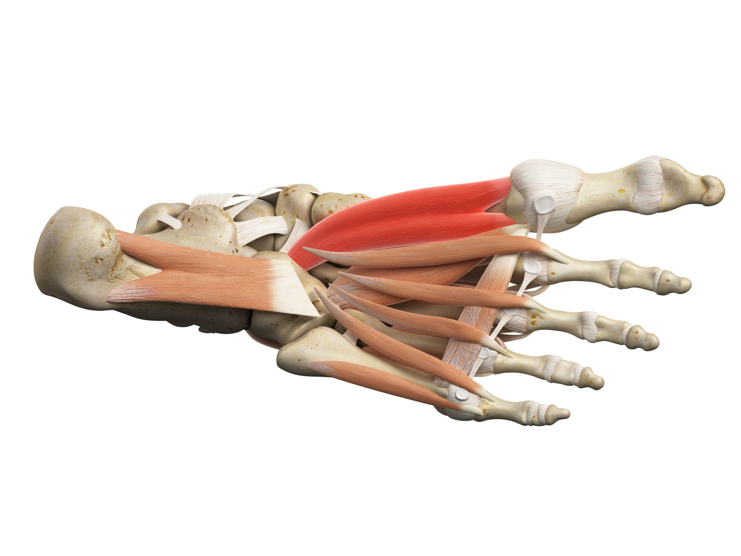
We could go on but how about you do some homework now?… why not research these little foot movers:

Delgado, Gonzalo J. et al (2002). “Tennis Leg: Clinical US Study of 141 Patients & Anatomic Investigation of Four Cadavers with MR Imaging & US1”. Radiology. 224 (1): 1129. doi:10.1148/radiol.2241011067. PMID 12091669
Freeman, A. J. et al (2008). “Anatomical variations of the plantaris muscle & a potential role in patellofemoral pain syndrome”. Clinical Anatomy. 21 (2): 178–81. doi:10.1002/ca.20594.PMID 18266282
Spina, A. A. (2007). “The plantaris muscle: Anatomy, injury, imaging, and treatment”. The Journal of the Canadian Chiropractic Association. 51 (3): 158–65. PMC 1978447. PMID 17885678
Do you feel like this is an area of the body you have yet to master?
Would you like a simple way of understanding & interpreting foot biomechanics to help more clients in more ways?